Describe the Steps for Preparing a Patient for Dental Imaging
The technologist will position you on the exam table and give you instructions. Dental patient preparation is the process of preparing a patient mentally and physically to receive dental care.
Pdf Dental Radiographic Procedures During Covid 19 Outbreak And Normalization Period Recommendations On Infection Control
Describe the methods used when preparing film for processing in an automatic processor.

. The direct method uses an electronic. The central ray of the x-ray beam is directed to intersect both the image receptor and the object tooth perpendicularly. The automatic processor see figure 3-3 is used by most dental treatment facilities.
Bisecting- angle instrument stabe bite-block and EeZee- Grip holder. The patients needs Step 2. 5 basic rules of paralleling technique.
Following exposure the film is unwrapped in the darkroom and immediately loaded into the automatic processor. Identify the patient-preparation and patient- positioning errors seen on panoramic images 7. Prepare for dental radiography.
Gloves removes hands cleaned patient dismissed clean area and take the films to the darkroom. The image isnt the same length as the tooth that is being x-rayed and is elonglated or foreshortened. The unit consists of rollers and compartments filled with chemical solutions through which the film advances.
Are There Any Risks of Having a Dental X-Ray. A few steps need to take place for most types of x-rays to prepare a patient. Describe how to prepare a patient for dental imaging.
What to Expect During an X-Ray. Dental patient preparation Definition. If all metal objects are not removed it will result in a ghost image if the lead apron is placed incorrectly a radiopaque cone-shaped artifact results if the patients lips are no closed a dark radiolucent shadow appear and obscures the anterior teeth if the chin is too high.
Describe the errors that occur during patient preparation and positioning during panoramic imaging. How to Prepare for an X-Ray. Image receptor must be placed away from the teeth and toward the middle of the mouth Explain why an image receptor holder is necessary with the paralleling technique.
Indirect digital imaging is a system that converts the traditional dental X-ray to digital imaging. Prepare the dental operatory. Dental X-rays radiographs are images of your teeth that your dentist uses to evaluate your oral health.
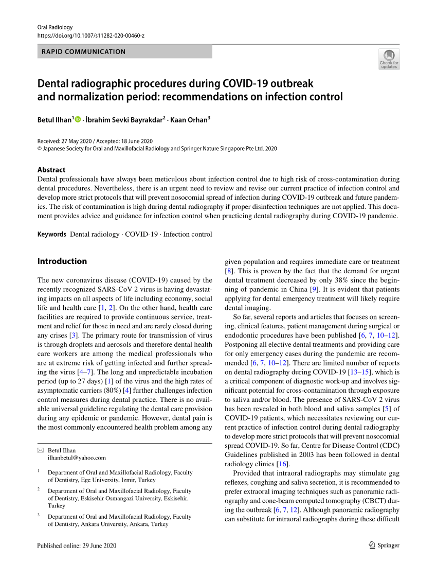
Patient comfort can be improved by placing digital receptors in a position that allows for accurate radiographs as well as using devices and accessories that improve patient comfort. To position the patient tell them to stand or sit with the back straight and erect instruct them to bite on the plastic bite lock position the midsagittal plane perpendicular to the floor position the Frankfort plane parallel to the floor and tell the patient to swallow saliva and keep the tongue on the roof of the mouth ask him or her to stay still and expose the film. Our 425-hour Dental Assistant program is divided into 4 in-depth courses and includes an additional 160-hour externship.
Patients can be protected through the use of lead collars and aprons and by ensuring that only necessary radiographs are taken and that radiation exposure is kept low. The most common approach to dental caries detection consists of dental radiography in conjunction with visual and tactile exploration. Prepared at least three patients at dental workplace for oral procedures.
Infection control practices for dental radiography are identical to those used in the operatory. Identify the types of image receptor holders that can be used with the bisecting technique. Describe patient preparation equipment preparation and patient- positioning procedures before exposing a panoramic projection.
Transfer from the dental chair to the wheelchair. Digital radiography is a type of X-ray imaging that uses digital X-ray sensors to replace traditional photographic X-ray film producing enhanced computer images of teeth gums and other oral structures and conditions. The technologist will verify your identification and exam requested.
Digital dental images are acquired through three methods. Each course covers a variety of topics including HIPAA rules and regulations endodontic procedures embryology dental imaging and so much more. Identify uses of dental radiography Describe the history of dental x-ray film List the properties of radiation Describe the radiation types Identify the radiation units of measurements Explain the biological effects of radiation exposure Identify the components of a dental x-ray unit and.
Position the patient after the transfer. Describe procedures during and after x-ray exposure. 7 At present however these routine procedures are not precise enough to detect early lesions especially in occlusal surfaces or gaps at the tooth-restoration interface which potentially lead to secondary caries.
12 Reassure the patient and explain the procedure in language that is easily understood. Describe the fundamentals of panoramic imaging 4. PPE dry exposed filmPSP when removing from patients mouth place in cup or black transfer box dispose of contaminated items wash hands and disinfect surface.
The direct method indirect method and semi-indirect method. 11 Position the patient comfortably. Completion will prepare you for the NELDA component exams and the RDA exam.
Describe the sequence of exposure for posterior teeth. Performance criteria describe the performance needed to demonstrate achievement of the element. Good preparation helps patients form a positive attitude about dental health care.
Ideally this attitude will continue throughout life enhancing optimal oral health. They are grounded in the practice of standard precautions and are directed toward preventing disease transmission from patients to DHCP from DHCP to patients from patient to patient and from the practice to the surrounding locale1-3. Ome patients who use wheelchairs can transfer themselves.
These X-rays are used with low levels of. The image receptor film packet phosphor plate or digital sensor is placed parallel to the long axis of the object tooth being radiographed. Describe the equipment used in panoramic imaging 5.
8 The early detection of. The duration of the exam will vary but the average is about 15 minutes. These steps include checking with female patients about a potential pregnancy having patients remove jewelry in the area that needs an x-ray and providing patients with protective gear such as a lead vest.
There are no special preparations for a diagnostic X-Ray exam.

Preparing The Patient For Dental Imaging By Karen Munoz

Patient Preparation Panoramic Radiographs Technique Anatomy Review Continuing Education Course Dentalcare Com

Procedures In Dental Imaging Pocket Dentistry

Dental Implants Procedure What You Need To Know Narre Warren Dental Care
Paralleling Technique In Dental Radiography
Patient Preparation Practical Panoramic Imaging Continuing Education Course Dentalcare Com
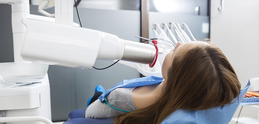
Dentistry Journal Free Full Text Maximizing Student Clinical Communication Skills In Dental Education Mdash A Narrative Review Html
What Is Fluoroscopy And How To Prepare Envision Radiology

Orthopantomography Radiology Reference Article Radiopaedia Org
What Is Fluoroscopy And How To Prepare Envision Radiology

Guidelines For Oral And Maxillofacial Imaging Covid 19 Considerations Oral Surgery Oral Medicine Oral Pathology And Oral Radiology

Children Free Full Text Restoration Of An Upper Anterior Tooth In An Adolescent With Autism Spectrum Disorder A Student Case Report Html

10 Steps To Efficient Endo In The General Practice Dental Economics
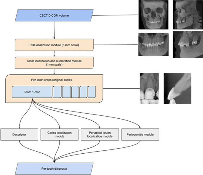
Clinically Applicable Artificial Intelligence System For Dental Diagnosis With Cbct Scientific Reports

Types Of Dental X Rays Are They Dangerous

Take It Right The First Time Registered Dental Hygienists

Preparing The Patient For Dental Imaging By Karen Munoz
Taking X Ray Examinations A Dental Assistant S Guide Meridian College

Virtual Diagnostics And Guided Tooth Preparation For The Minimally Invasive Rehabilitation Of A Patient With Extensive Tooth Wear A Validation Of A Digital Workflow Journal Of Prosthetic Dentistry


Comments
Post a Comment